Showing posts with label Pituitary tumors. Show all posts

Monday, February 23, 2015

Top Endocrine Publications of 2014: The Canine and Feline Pituitary Gland


For my next review of the endocrine publications of 2014 that concern companion animals, I'm going to turn to the theme of diagnosis and treatment of pituitary problems in dogs and cats. Listed below are 18 clinical and research papers written in 2014 that deal with a variety of pituitary gland issues of clinical importance in dogs and cats.

These range from case studies of cats with primary hypodipsia and inappropriate antidiuretic hormone secretion (1,2) to an investigation of the clinical utility of formulas of estimated serum osmolality (3); from a study of acromegaly in a series German shepherd dogs (4) to a number of excellent studies of the clinical features, diagnosis, or treatment of feline acromegaly (8,9,13,15); and from investigation of the stress response in dogs (5,14) to a study of the intraoperative changes of circulating vasopressin during elective ovariohysterectomy in dogs (6).

Other publications include a study investigating the problems associated with commercial assays for determination of feline ACTH (7) to a review of the use of GnRH agonists in dogs and cats (10); from a report of a transsphenoidal surgical technique for removal of pituitary adenomas in dogs with pituitary-dependent Cushing's disease (11) to a review of the role of prolactin in canine mammary tumor development (12); and finally, from a report of the clinical findings, diagnostic test results, and treatment outcome of 30 cats with spontaneous Cushing's disease (16) to an investigation of the mutations associated with pituitary dwarfism in Saarloos and Czechoslovakian wolfdogs (18).

References:
  1. Bach J, Claus K. Primary hypodipsia in a cat with severe hypernatremia. J Feline Med Surg 2014;16:240-242. 
  2. Demonaco SM, Koch MW, Southard TL. Syndrome of inappropriate antidiuretic hormone secretion in a cat with a putative Rathke's cleft cyst. J Feline Med Surg 2014;16:1010-1015. 
  3. Dugger DT, Epstein SE, Hopper K, et al. A comparison of the clinical utility of several published formulae for estimated osmolality of canine serum. J Vet Emerg Crit Care (San Antonio) 2014;24:188-193. 
  4. Fracassi F, Zagnoli L, Rosenberg D, et al. Spontaneous acromegaly: a retrospective case control study in German shepherd dogs. Vet J 2014;202:69-75. 
  5. Hekman JP, Karas AZ, Sharp CR. Psychogenic stress in hospitalized dogs: cross species comparisons, implications for health care, and the challenges of evaluation. Animals (Basel) 2014;4:331-347. 
  6. Hoglund OV, Hagman R, Olsson K, et al. Intraoperative changes in blood pressure, heart rate, plasma vasopressin, and urinary noradrenalin during elective ovariohysterectomy in dogs: repeatability at removal of the 1st and 2nd ovary. Vet Surg 2014;43:852-859. 
  7. Kemppainen RJ. Amino acid differences in cat adrenocorticotropin account for the inability of a human-based immunoradiometric assay to detect the molecule in cat plasma. J Vet Diagn Invest 2014;26:431-433.
  8. Lamb CR, Ciasca TC, Mantis P, et al. Computed tomographic signs of acromegaly in 68 diabetic cats with hypersomatotropism. J Feline Med Surg 2014;16:99-108. 
  9. Lourenco BN, Randall E, Seiler G, et al. Abdominal ultrasonographic findings in acromegalic cats. J Feline Med Surg 2014.  
  10. Lucas X. Clinical use of deslorelin (GnRH agonist) in companion animals: a review. Reprod Domest Anim 2014;49 Suppl 4:64-71. 
  11. Mamelak AN, Owen TJ, Bruyette D. Transsphenoidal surgery using a high definition video telescope for pituitary adenomas in dogs with pituitary dependent hypercortisolism: methods and results. Vet Surg 2014;43:369-379. 
  12. Michel E, Rohrer Bley C, Kowalewski MP, et al. Prolactin--to be reconsidered in canine mammary tumourigenesis? Vet Comp Oncol 2014;12:93-105. 
  13. Myers JA, Lunn KF, Bright JM. Echocardiographic findings in 11 cats with acromegaly. J Vet Intern Med 2014;28:1235-1238. 
  14. Nagasawa M, Shibata Y, Yonezawa A, et al. The behavioral and endocrinological development of stress response in dogs. Dev Psychobiol 2014;56:726-733. 
  15. Rosca M, Forcada Y, Solcan G, et al. Screening diabetic cats for hypersomatotropism: performance of an enzyme-linked immunosorbent assay for insulin-like growth factor 1. J Feline Med Surg 2014;16:82-88. 
  16. Valentin SY, Cortright CC, Nelson RW, et al. Clinical findings, diagnostic test results, and treatment outcome in cats with spontaneous hyperadrenocorticism: 30 cases. J Vet Intern Med 2014;28:481-487. 
  17. van Rijn SJ, Riemers FM, van den Heuvel D, et al. Expression stability of reference genes for quantitative RT-PCR of healthy and diseased pituitary tissue samples varies between humans, mice, and dogs. Mol Neurobiol 2014;49:893-899. 
  18. Voorbij AM, Leegwater PA, Kooistra HS. Pituitary dwarfism in Saarloos and Czechoslovakian wolfdogs is associated with a mutation in LHX3. J Vet Intern Med 2014;28:1770-1774. 

Wednesday, February 19, 2014

Top Endocrine Publications of 2013: The Canine and Feline Pituitary Gland


As I've done for the last four years, I’ve now finished compiling a fairly extensive list of references concerning canine and feline endocrinology that were written last year (in 2013). I’ll be sharing these with you over the next few weeks, as well as reviewing a few of the best papers from my lists of clinical endocrine publications.

In this post, I am going to start off with papers that deal with the theme of diagnosis and treatment of pituitary problems in dogs and cats.

Listed below are 13 clinical and research papers written in 2013 that deal with a variety of pituitary gland issues of clinical importance in dogs and cats.

These range from studies of the pathogenesis of acromegaly (and diabetes) in cats (2) to two excellent reviews of the clinical features, diagnosis, and treatment of feline acromegaly (8,9); from a case report of a cat with pituitary adenomas secreting both ACTH and GH (12) to another case report of a cat suffering from a pituitary carcinoma causing hyperadrenocorticism (6); and from a study of the accuracy of CT and MRI for contouring the feline apparatus for radiation therapy planning (for treatment of feline acromegaly) (10) to studies validating an assay for feline ACTH determination (3).

Other publications include a case report of two dogs that presented with severe polyuria and polydipsia due to thyroid carcinoma and hyperthyroidism (1) to diabetes insipidus (DI) in a cat secondary to head trauma (11); and a report on acute iatrogenic water intoxication in cats (7) to a study of the disturbances of water metabolism (normovolemic hypernatremia) secondary to pituitary gland/hypothalamic dysfunction (13).

References:
  1. Bosje T, den Hertog E, Dijksta M. Does the T4 measurement belong in the standard blood analysis in polyuria/polydipsia? Tijdschr Diergeneeskd 2013;138:230-231. 
  2. Dirtu AC, Niessen SJ, Jorens PG, et al. Organohalogenated contaminants in domestic cats' plasma in relation to spontaneous acromegaly and type 2 diabetes mellitus: A clue for endocrine disruption in humans? Environ Int 2013;57-58:60-67. 
  3. Eiler KC, Bruyette DS, Behrend EN, et al. Comparison of intravenous versus intramuscular administration of corticotropin-releasing hormone in healthy cats. J Vet Intern Med 2013;27: 516-521. 
  4. Frischknecht M, Niehof-Oellers H, Jagannathan V, et al. A COL11A2 mutation in Labrador retrievers with mild disproportionate dwarfism. PLoS One 2013;8:e60149. 
  5. Goericke-Pesch S, Georgiev P, Fasulkov I, et al. Basal testosterone concentrations after the application of a slow-release GnRH agonist implant are associated with a loss of response to buserelin, a short-term GnRH agonist, in the tom cat. Theriogenology 2013;80:65-69. 
  6. Kimitsuki K, Boonsriroj H, Kojima D, et al. A case report of feline pituitary carcinoma with hypercortisolism. J Vet Med Sci 2014;76:133-138. 
  7. Lee JY, Rozanski E, Anastasio M, et al. Iatrogenic water intoxication in two cats. J Vet Emerg Crit Care (San Antonio) 2013;23:53-57. 
  8. Niessen SJ. Update on feline acromegaly. In Practice 2013;35:2-6. 
  9. Niessen SJ, Church DB, Forcada Y. Hypersomatotropism, acromegaly, and hyperadrenocorticism and feline diabetes mellitus. Vet Clin North Am Small Anim Pract 2013;43:319-350. 
  10. Nolan MW, Randall EK, LaRue SM, et al. Accuracy of CT and MRI for contouring the feline optic apparatus for radiation therapy planning. Vet Radiol Ultrasound 2013;54:560-566. 
  11. Oliveira KM, Fukushima FB, Oliveira CM, et al. Head trauma as a possible cause of central diabetes insipidus in a catJ Feline Med Surg 2013;15:155-159. 
  12. Sharman M, FitzGerald L, Kiupel M. Concurrent somatotroph and plurihormonal pituitary adenomas in a catJ Feline Med Surg 2013;15:945-952. 
  13. Weingart A, Gruber AD, Kershaw O, et al. Disturbances of water metabolism in two dogs and one cat with central nervous system disorders. Schweiz Arch Tierheilkd 2013;155:463-469. 

Tuesday, July 2, 2013

Top 10 Clinical Endocrinology Research Abstracts Presented at the 2013 ACVIM Meeting

Last month, I spent a week in Seattle, Washington attending the the 2013 American College of Veterinary Internal Medicine Forum.  As part of that meeting, a number of research abstracts were presented (oral and poster presentations) that dealt with various aspects of canine and feline endocrinology. I plan to spend the next three blogs discussing some of the newest and best research findings featured at the ACVIM meeting.

Of all of the excellent endocrine research abstracts presented, I've selected a "top 10 list" of the ones that have the most potential to change what I do in my clinical practice.  To do this, I've enlisted the help of Dr. Rhett Nichols, a well-known expert in endocrinology and internal medicine whose day-job is senor member of the veterinarian consulting service for Antech Diagnostics, the world's largest laboratory dedicated to animal health.  However, since Rhett also serves as a consultant for the Animal Endocrine Clinic (my practice), it was not that difficult to get him involved in this project!

In this post, we will review 4 of these top 10 abstracts, followed by the remaining 6 in the upcoming 2 posts. We hope you agree with our selections, but if you don't, remember that you can always post a comment and add your opinion.

Niessen S, Scudder C, Forcada Y, et al. Pasireotide (SOM230) opens doors to medical management of feline hypersomatotropism. J Vet Intern Med 2013:685.

Feline hypersomatotropism (HS) appears to be a significant cause of feline diabetes mellitus. However, successful treatment of HS is currently challenging. Radiotherapy and hypophysectomy seem the only effective therapeutic modalities, yet come with significant disadvantages. Medical options would be desirable although somatostatin (sst) analogues and dopamine agonists have thus far proven largely ineffective. Pasireotide (SOM230), a novel multi-receptor ligand sst analogue with high binding affinity for sst receptor subtypes 1, 2, 3 and 5 has been shown to suppress growth hormone (GH) and insulin-like growth factor-1 (IGF-1) in rodents as well as humans suðering from HS. Additionally, direct and indirect anti-tumor activity has been observed in vitro including sst receptor-mediated apoptosis and anti-angiogenesis. The current study aimed to assess the potential of SOM230 as a treatment modality for naturally occurring feline HS. Feline HS was diagnosed in eight diabetic cats by documenting serum IGF-1 concentration >1000 ng/ml (radioimmunoassay) and presence of a pituitary enlargement (computed tomography). On day 1 and 5, serum IGF-1 concentration was established and glycemic control assessed using a 12-hour blood glucose (BG) curve, measuring BG every 2 hours. On day 2, 3 and 4, the cats were injected with 0.03 mg/kg SOM230 s.c. BID. The initial insulin dose was dictated by the choice of the attending clinician, although was reduced according to regular BG measurements during the treatment period to avoid hypoglycemia. Pre- and post-treatment IGF-1, average 12-hour BG and insulin dose were compared using a paired t-test (significance at P < 0.05). All eight cats showed a significant decrease in serum IGF-1 (mean+/-SD day 1: 1884 + /-218 ng/ml; day 5: 1169 + /-395 ng/ ml, p = 0.001) and average 12-hour BG (day 1: 20 + /-5 mmol/l; day 5: 13 + /-4 mmol/l; p = 0.002). A significant insulin dose reduction was necessary in all cats (day 1: 10.8 + /-6 iu/injection; day 5: 3.1 + /-2 iu/injection; p = 0.015). No side effects were noticed during or after the 3 day treatment period, apart from hypoglycemia in one cat, which resolved after provision of food and reduction of insulin dose. The current study indicates that SOM230 is able to rapidly decrease GH and IGF-1 concentrations in feline HS. This, therefore, suggests that sst receptors are present in most feline somatotrophinomas, which has previously been unclear given the disappointing results during somatostatins and sst analogue therapy attempts. A return of insulin sensitivity was seen, enabling improved glycemic control to be established with reduced doses of exogenous insulin in all cats. In light of these results, a clinical trial with a longer-acting formulation of SOM230 is currently being conducted to establish long-term effects and potential for diabetic remission. 

Comments— Pasireotide (SOM230, trade name Signofor, Novartis) is an orphan drug approved for the treatment of Cushing’s disease in adult human patients when surgery has failed or is not an option (1). The drug is a somatostatin analog that targets multiple somatostatin receptors with high affinity. The result is apoptosis of those cells that produce ACTH, with significant lowering of plasma ACTH levels (2,3).

In addition, pasireotide has been shown to suppress GH and IGF -1 in rodents and human patients with acromegaly (4). Moreover, recent results of a phase III study of human patients with acromegaly treated with a long-acting release form of pasireotide show that this novel form of therapy is significantly more effective than the current standard therapy with octreotide (5).

This study by Niessen et al indicates that pasireotide is able to rapidly decrease GH and IGF-1 concentrations in feline acromegaly and suggests that somatostain receptors are present in most cats with pituitary tumors that produce excessive GH. In light of these results, a clinical trial with the long-acting release form of pasireotide is currently being conducted to establish long-term effects and potential for diabetic remission in cats with acromegaly.

The Bottom Line—It is great to finally have a medical treatment that may actually work for cats with acromegaly. Unfortunately, administration of pasireotide SC twice daily may not be a practical or affordable therapeutic option for many of our cat owners.

References:
  1. Signifor Official Site - Signifor® (pasireotide) Injection. Signifor.US‎. 
  2. Colao A, Petersenn S, Newell-Price J, et al. A 12-month phase 3 study of pasireotide in Cushing's disease. N Engl J Med 2012;366:914-924. 
  3. McKeage K. Pasireotide: a review of its use in Cushing's disease. Drugs 2013;73:563-574. 
  4. Petersenn S, Farrall AJ, Block C, et al. Long-term efficacy and safety of subcutaneous pasireotide in acromegaly: results from an open-ended, multicenter, Phase II extension study. Pituitary 2013. DOI 10.1007/s11102-013-0478-0 
  5. Colao A, Bronstein M, Freda P, et al. Pasireotide LAR is significantly more effective than octreotide LAR at inducing biochemical control in patients with acromegaly: Results of a 12-month randomized, double-blind, multicenter, Phase III study. Joint 15th International Congress of Endocrinology and 14th European Congress of Endocrinology. Abstract #OC1.1. 2012 

De Marco V, Noronha KSM, Casado TC, et al. Therapy of canine hyperlipidemia with bezafibrate. J Vet Intern Med2013;27:694.

The primary and secondary hyperlipidemia are common in dogs and its treatment is necessary to prevent clinical complications such as pancreatitis, seizures, liver disease and diabetes. The therapy of mild hyperlipidemia comprising a fat restricted diet, but in more severe cases pharmacological treatment is necessary. Bezafibrate (BZF) is effective in the treatment of hypertriglyceridemia in humans, however there are no clinical studies in dogs. The objectives of this study were to assess the efficacy of BZF in reducing serum triglyceride (TG) and cholesterol (CHO) in hyperlipidemic dogs, identify a therapeutic protocol for this drug and assess possible side eðects such as muscle pain, emesis, diarrhea and elevated CK and TGP levels. Only animals with moderate to severe hypertriglyceridemia (TG> 350 mg/dL) were treated with BZF every 24 hours for 30 days before introduction of any other therapy according to the protocol: tablet 200 mg for dogs weighting less than 12 kg, tablet 200 mg for dogs weighing between 13 and 25 kg, 1 tablet 200 mg for dogs weighing over 25 kg. We studied 46 dogs (26 females and 20 males) with a mean age of 9 years. Fifteen dogs (32.6%) had primary hyperlipidemia and 31 (67.4%) secondary hyperlipidemia, which included hyperadrenocorticism (41.3%), hypothyroidism (15.2%) and chronic corticoideterapia (10.8%). All 46 (100%) dogs had hypertriglyceridaemia and 33 (71.7%) had both hypertriglyceridaemia and hypercholesterolemia. After 30 days using BZF, normalization of serum TG (TG <150 mg/dL) was observed in 91.3% of cases (n = 42/46) and of CHO (CHO < 270 mg/dL) in 66 7% (n = 22/33) of cases. Means and standard deviations of serum TG and COL before (752 ± 663 mg/dL and 428 ± 217 mg/dL) and after therapy (110 ± 82 and 244 ± 71 mg /dL) were significantly lower (p < 0.005, paired Student t test). The bezafibrate dose most used with a 95% confidence interval was 5.3 to 6.1 mg/kg (range: 4–10 mg/kg). No side effects were observed, and there was no statistical difference between the values of ALT and CK before and after therapy. It can be concluded that bezafibrate is a safe and effective drug for the canine hyperlipidemia therapy.
  
Comments—Bezafibrate is a fibrate drug used for the treatment of hyperlipidemia (1-3). In people, fibrates are used as an accessory drug in many forms of hypercholesterolemia, usually along with statins. Bezafibrate helps lower cholesterol and triglycerides in the blood and increase high density lipoproteins (HDL). The main toxicity is hepatic, myopathy, and rarely rhabomyolysis.

Hyperlipidemia is a relatively commonly recognized disorder in dogs but management can be frustrating (4). In this study, 46 dogs with primary or secondary hyperlipidemia (diabetes mellitus, Cushing’s syndrome. hypothyroidism) were treated with bezafibrate once a day over a 30-day period; triglycerides and cholesterol were significantly lowered in the majority of dogs. In addition, there was no evidence of untoward side effects (e.g., no clinical issues and ALT and CK levels were not altered).

There are 2 preparations of bezafibrate available: 200 mg tablets and 400 mg sustained-release tablets. The sustained-release preparation is taken once a day; the non-sustained release tablets are taken with each meal. For dogs, the average dose used in this study was 5 to 6 mg/kg once a day. The dosing protocol was ¼ of a 200 mg tablet for dogs < 12 kg, ½ of a 200 mg tablet for dogs weighing between 12 and 25 kg, and one 200 mg tablet for dogs > 25 kg.

The Bottom Line—Bezafibrate given once a day appears to be a safe and effective drug for the treatment of hyperlipidemia in the dog.

References:
  1. Goa KL, Barradell LB, Plosker GL. Bezafibrate. An update of its pharmacology and use in the management of dyslipidaemia. Drugs 1996;52:725-753.  
  2. Goldenberg I, Benderly M, Goldbourt U. Update on the use of fibrates: focus on bezafibrate. Vasc Health Risk Manag 2008;4:131-141.  
  3. Teramoto T, Shirai K, Daida H, et al. Effects of bezafibrate on lipid and glucose metabolism in dyslipidemic patients with diabetes: the J-BENEFIT study. Cardiovasc Diabetol 2012;11:29. 
  4. Xenoulis PG, Steiner JM. Lipid metabolism and hyperlipidemia in dogs. Vet J 2010;183:12-21.

Salesov E, Boretti FS, Sieber-Ruckstuhl NS, et al. Urinary and plasma catecholamine and metanephrine in dogs with pheochromocytoma, hyperadrenocorticism and in healthy dogs. J Vet Intern Med 2013 27:688-689.

Pheochromocytoma (PHEO) is a rare malignant catecholamine-secreting tumor of the adrenal medulla. Catecholamines and metanephrines in plasma and in 24-h urine are approved biomarkers for the detection of the disease in humans, however, the question which of the tests is best is controversial. We previously demonstrated that measurement of urinary catecholamine and metanephrine to creatinine ratios is helpful for the diagnosis of PHEO in dogs and that urinary normetanephrine to creatinine ratio may be the best test to discriminate between PHEO and hypercortisolism (HC). Knowledge on plasma catecholamines and metanephrines in dogs is scarce and no comparison between urinary and plasma parameters has been performed. The objective of the study was to measure urinary as well as plasma catecholamines and metanephrines in dogs with PHEO, HC and in healthy dogs and to determine the test with the least overlap between the group. Six dogs with PHEO, 9 dogs with HC (6 with ATH, 3 with PDH) and 10 healthy dogs were included. Urine samples were collected into HCL containing tubes to ensure a pH 2, blood samples were collected on ice, centrifuged at 4°C and immediately snap frozen in liquid nitrogen. All samples were stored at – 80°C. Urinary epinephrine (U-Epi), norepinephrine (U-Norepi), metanephrine (U-Meta) and normetanephrine (U-Normeta), and epinephrine (P-Epi), norepinephrine (P-Norepi), free and total metanephrine (PF-Meta and PT-Meta) and free and total normetanephrine (PF-Normeta and PT-Meta) were analysed by HPLC. Urinary catecholamines and metanephrines were expressed as ratios to urine creatinine concentrations. Data were analysed by non-parametric tests (P < 0,05). Similar to our previous findings U-Epi, U-Norepi, U-Meta and U-Normeta were significantly higher in dogs with PHEO and U-Norepi and U-Normeta were significantly higher in dogs with HC compared to healthy dogs. Comparison between dogs with HC and dogs with PHEO revealed significantly higher U-Meta and U-Normeta in the latter group. U-Normeta was the only parameter with no overlap. In dogs with PHEO P-Norepi, PF-Meta, PT-Meta, PF-Normeta, PT-Normeta were significantly higher and in dogs with HC P-Norepi, PF- Normeta and PT-Normeta were significantly higher than in healthy dogs. Comparison between dogs with HC and dogs with PHEO showed significant higher PF-Meta, PT-Meta, PF- Normeta, PT-Normeta in the PHEO group. Overlap was present with all 4 parameters, but was least with PF-Normeta and PT-Normeta. According to our results U-Normeta, PF- Normeta and PT-Normeta are valuable parameters for the diagnosis of PHEO, so far U-Normeta performed better than the plasma parameters. 

Comments—In some recent studies, up to one in five adrenal tumors has been a pheochromocytoma. In the past, a presumptive diagnosis of a pheochromocytoma was based on history which was often a vague, sometimes episodic description of illness, documentation of hypertension, an adrenal mass noted on abdominal ultrasound, and ruling out adrenal-dependent Cushing’s syndrome with an endogenous ACTH level or the results of dexamethasone suppression testing. This current research adds additional data to the idea that measurement of urinary and plasma catecholamine and metanephrine can also be used to aid in diagnosis.

The Bottom Line—Currently, a urine normetaphrine/creatinine level, appears to be the most sensitive and specific test to document a pheochromocytoma.  This test requires that the urine sample is acidified at the time of collection and a control urine sample from a normal dog (also acidified) is submitted. A urine normetaphrine/creatinine level at least 4-times the control is consistent with pheochromocytoma.

In the US, the test for urine normetaphrine/creatinine can be performed at Marshfield Labs (www.marshfieldlabs.com). Acid pellets for urinary acidification are available from the laboratory.

References:
  1. Quante S, Boretti FS, Kook PH, et al. Urinary catecholamine and metanephrine to creatinine ratios in dogs with hyperadrenocorticism or pheochromocytoma, and in healthy dogs. J Vet Intern Med 2010;24:1093-1097. 
  2. Kook PH, Grest P, Quante S, et al. Urinary catecholamine and metadrenaline to creatinine ratios in dogs with a phaeochromocytoma. Vet Rec2010;166:169-174. 
  3. Kook PH, Boretti FS, Hersberger M, et al. Urinary catecholamine and metanephrine to creatinine ratios in healthy dogs at home and in a hospital environment and in 2 dogs with pheochromocytoma. J Vet Intern Med2007;21:388-393. 

Sangster K, Panciera JL, Abbott A, et al. Cardiac biomarkers in hyperthyroid cats. J Vet Intern Med 2013:637. 

Differentiation of hyperthyroid heart disease from primary myocardial disease is challenging. The cardiac biomarkers NT- proBNP and troponin I (cTNI) have proven useful in identifying cats with myocardial disease and may provide a method by which hypertrophic cardiomyopathy (HCM) and hyperthyroid heart disease can be discriminated. The primary purpose of this study was to compare plasma concentrations of NT-proBNP and cTNI in three groups of cats: cats with naturally occurring hyperthyroidism, cats with primary cardiomyopathy, and healthy older cats to determine if biomarkers differ between groups and if bio-marker concentrations in hyperthyroid cats change after resolution of the thyroid disease. We prospectively evaluated 61 client-owned cats: 23 hyperthyroid cats, 19 cats with HCM without congestive heart failure, and 19 euthyroid, normotensive healthy cats eight years of age or older. Fourteen of the hyperthyroid cats were re-evaluated three months after administration of I-131. A complete history, physical examination, CBC, serum biochemistries, urinalysis, blood pressure measurement, serum T4 concentration, plasma concentrations of NT-proBNP and cardiac troponin I, and echocardiography was obtained for each cat. Hyperthyroid and HCM cats had plasma NT-proBNP and cTNI concentrations that were significantly greater than healthy older cats, but there was no significant difference between hyperthyroid and HCM cats with respect to concentration of either biomarker. Plasma NT-proBNP and cTNI concentrations decreased in each cat that was examined three months after I-131 treatment. Plasma cTNI was within the reference interval for all cats at the three month recheck. Severely thickened myocardium persisted in one formerly hyperthyroid cat at the three month recheck, and this cat’s plasma NT-proBNP remained elevated. Although there may be a role for NT-proBNP in monitoring the cardiac response to treatment of hyperthyroidism, neither NT-proBNP nor cTNI can be used to distinguish hyperthyroid cats from cats with HCM. Therefore, the thyroid status of older cats should be ascertained prior to interpreting results of cardiac biomarker testing.
  
 Comments—Although it is well established that hyperthyroid cats will commonly develop secondary heart disease (1), it can sometimes be difficult to distinguish thyroid-induced cardiac disease from primary myocardial disease (cardiomyopathy). Over the last few years, a number of studies have confirmed the usefulness of plasma cardiac biomarkers (especially N-terminal pro-brain natriuretic peptide or NT-proBNP) to help detect hypertrophic cardiomyopathy in cats and to distinguish primary cardiac from non-cardiac causes of dyspnea in cats (2-5). Previous studies have found that hyperthyroid cats can have high circulaing levels of either troponin I or NT-proBNP; both biomarkers fall after successful treatment of the hyperthyroid state (6,7).

This research study confirmed that hyperthyroid cats can have high plasma NT-proBNP and troponin I (cTNI) concentrations, which decreased after I-131 treatment. However, there was no significant difference between hyperthyroid and HCM cats with respect to concentration of either biomarker.

The Bottom Line— Although hyperthyroid cats can have high plasma NT-proBNP and cTNI concentrations, there was no significant difference between hyperthyroid and HCM cats with respect to concentration of either biomarker. Therefore, neither of these cardiac biomarkers can be used to distinguish hyperthyroid cats from cats with HCM. Since hyperthyroidism can result in high levels of both biomarkers (6,7), the thyroid status of older cats should always be ascertained prior to interpreting results of cardiac biomarker testing.

References:
  1. Syme HM. Cardiovascular and renal manifestations of hyperthyroidism. Vet Clin North Am Small Anim Pract 2007;37:723-743, vi. 
  2. Wells SM, Sleeper M. Cardiac troponins. J Vet Emerg Crit Care 2008;18:235–245. 
  3. Boswood A. Biomarkers in cardiovascular disease: beyond natriuretic peptides. J Vet Cardiol 2009;11 Suppl 1:S23-32. 
  4. Fox PR, Oyama MA, Reynolds C, et al. Utility of plasma N-terminal pro-brain natriuretic peptide (NT-proBNP) to distinguish between congestive heart failure and non-cardiac causes of acute dyspnea in cats. J Vet Cardiol 2009;11 Suppl 1:S51-61. 
  5. Wess G, Daisenberger P, Mahling M, et al. Utility of measuring plasma N-terminal pro-brain natriuretic peptide in detecting hypertrophic cardiomyopathy and differentiating grades of severity in cats. Vet Clin Pathol 2011;40:237-244. 
  6. Connolly DJ, Guitian J, Boswood A, et al. Serum troponin I levels in hyperthyroid cats before and after treatment with radioactive iodine. J Feline Med Surg 2005;7:289-300. 
  7. Menaut P, Connolly DJ, Volk A, et al. Circulating natriuretic peptide concentrations in hyperthyroid cats. J Small Anim Pract 2012;53:673-678.  

Monday, September 10, 2012

Pituitary MRI Imaging in Cats with Acromegaly


PAPER REVIEW

Magnetic Resonance Imaging Findings in 15 Acromegalic Cats
by Barbara Posch, Jane Dobson, Mike Herrtage
Veterinary Radiology & Ultrasound 2011;52:422–427.

Background
MRI imaging of a cat with acromegaly.
Note the large pituitary mass that has
expanded dorsally into the hypothalamus
In cats, chronic hypersecretion of growth hormone (GH) causes acromegaly, a disease characterized by insulin-resistant diabetes mellitus and progressive overgrowth of soft tissue, membranous bone, and viscera (1-3). Acromegaly has been considered rare in cats but it may be more common than suspected previously (4-6). In almost all cats, the cause of acromegaly is a GH-secreting pituitary tumor (1-7).

Definitive diagnosis of feline acromegaly can be difficult because of the gradual onset and subtle clinical signs and unavailability of a validated feline GH assay (2,3). Therefore, the diagnosis of acromegaly in cats is currently based upon a combination of clinical signs, the finding of high serum insulin-like growth factor-1 (IGF-1) concentrations, and the documentation of a pituitary mass on brain imaging (2-6).

The usefulness of  computed tomography (CT) and magnetic resonance imaging (MRI) in identifying a pituitary tumor and establishing a diagnosis of acromegaly in cats has been demonstrated in many reports (4,5,7). However, little information is available regarding MRI features of pituitary tumors in acromegalic cats.

Objectives
The purpose of this study by Posch et al (8) was to evaluate if pituitary abnormalities were present on MRI imaging in acromegalic cats and to determine if specific morphologic criteria can be established to aid in diagnosis.

Animals
Fifteen cats with acromegaly were included in this study.

Methods
Retrospective study. Inclusion criteria included the following: 1) high serum IGF-1 value (>100 ng/ml); 2) insulin-resistant diabetes mellitus (>1.5 U/kg per injection); 3) high serum fructosamine; 4) continued signs of diabetes (polyuria, polydipsia, polyphagia) despite insulin treatment; and 5) exclusion of other causes of insulin resistance.

MRI of the brain was performed using a 0.2 T scanner with a dual-phased array coil. T2-weighted  and pre- and post-contrast T1-weighted images were acquired in transverse and sagittal planes. Contrast-enhanced T1-weighted images were obtained following an intravenous bolus of 0.1 mmol/kg of gadobenate dimeglumine. Fluid attenuation inversion recovery (FLAIR) images were acquired in transverse plane in four cats.

The pituitary gland was measured on postcontrast T1-weighted images. Signal intensity was described relative to the normal cerebral cortical gray matter. Suprasellar extension, compression of the hypothalamus, dorsal displacement of the third ventricle were recorded.

Results
Enlargement of the pituitary gland with suprasellar extension was present in all 15 acromegalic cats. No characteristic signal patterns were identified on T1-weighted and T2-weighted sequences.

Contrast enhancement was nonuniform in all cats, as was suspected involvement of the adjacent hypothalamus. A mass effect on the cavernous sinus and third ventricle was present in 13 of the 15 cats. Mild peritumoral edema was present in four cats, and moderate edema in one cat. Transtentorial herniation was present in one cat.

Histopathology confirmed the presence of a pituitary adenoma in two cases.

Clinical conclusions
MRI is a useful modality to establish the diagnosis of acromegaly. Large pituitary masses were present in all cats but there was no consistent morphologic criterion. In most cats, the pituitary tumor had a heterogeneous appearance on both T1-weighted and T2-weighted images, with nonuniform contrast enhancement.

My Bottom Line:

Published reports of acromegaly in cats are relatively sparse but have been gradually increasing since the disease was first described 30 years ago (1,6). Although thought to be a rare disorder by most veterinarians, recent research suggests that the prevalence underlying acromegaly in cats with diabetes may actually be as high as 20 to 30% (4-6). This strongly suggests that this disorder is greatly underdiagnosed by practicing veterinarians today.

Because of the limited availability of a validated growth hormone assay in cats, measurement of IGF-1 is used as a screening test for acromegaly (2-5). For the practicing veterinarian, IGF-1 determinations are widely available and are performed by most commercial veterinary laboratories, in contrast to the limited availability of feline GH assays.

The basis for use of IGF-1 as a diagnostic test for acromegaly is that circulating GH activates the hepatic and peripheral tissue production of IGF-1, which is responsible for many of the actions normally attributable to GH itself. In fact, other than DM, most of the clinical features of acromegaly are not the result of a direct catabolic effect of GH excess, but rather result from an indirect anabolic effect of GH excess mediated through the production of the IGF-1 (e.g., overgrowth of soft tissue, bony enlargement, and organomegaly). Therefore circulating IGF-1 levels serve as a biomarker for assessing the peripheral biological effect of GH hypersecretion, which, at least in human patients, tends to correlate better with the severity of the acromegalic state than does random circulating GH determinations (9).

Although measurement of IGF-1 levels are useful in making a diagnosis of feline acromegaly, this is not a perfect diagnostic test. False-positive (and false-negative) results do occur, and high serum concentrations of serum IGF-1 is not uncommon in diabetic cats without acromegaly (10,11).  For that reason, the definitive diagnosis of acromegaly is best made by measuring a high IGF-1 combined with the finding of a pituitary mass with brain imaging (either MRI or CT).

It appears that MRI may be a more sensitive diagnostic test for this condition (2,3). However, virtually all cats with acromegaly have a clearly visible pituitary tumor on CT or MRI imaging. Therefore, at least clinically, it does not appear to make a large difference which imaging technique is chosen.

References:
  1. Peterson ME, Taylor RS, Greco DS, et al. Acromegaly in 14 cats. Journal of Veterinary Internal Medicine 1990;4:192–201.
  2. Niessen S, Peterson ME, Church DB. Acromegaly In: Mooney CT,Peterson ME, eds. BSAVA Manual of Canine and Feline Endocrinology. Fourth ed. Quedgeley, Gloucester: British Small Animal Veterinary Association, 2012;35-42.
  3. Peterson ME. Pituitary disorders In: Little SE, ed. The Cat: Clinical Medicine and Management. St. Louis: Elsevier Saunders, 2012;610-625.
  4. Niessen SJ, Petrie G, Gaudiano F, et al. Feline acromegaly: an underdiagnosed endocrinopathy? Journal of Veterinary Internal Medicine 2007;21:899–905.
  5. Berg RI, Nelson RW, Feldman EC, et al. Serum insulin-like growth factor-I concentration in cats with diabetes mellitus and acromegaly. Journal of Veterinary Internal Medicine 2007;21:892–898.
  6. Peterson ME. Acromegaly in cats: are we only diagnosing the tip of the iceberg? Journal of Veterinary Internal Medicine 2007;21:889–891.
  7. Dunning MD, Lowrie CS, Bexfield NH, Dobson JM, Herrtage ME. Exogenous insulin treatment after hypofractionated radiotherapy in cats with diabetes mellitus and acromegaly. Journal of Veterinary Internal Medicine 2009;23:243–249.
  8. Melmed S. Acromegaly pathogenesis and treatment. Journal of Clinical Investigation 2009; 119:3189-3202.
  9. Posch B, Dobson J, Herrtage M. Magnetic resonance imaging findings in 15 acromegalic cats. Veterinary Radiology & Ultrasound 2011;52:422–427.
  10. Lewitt MS, Hazel SJ, Church DB, et al. Regulation of insulin- like growth factor-binding protein-3 ternary complex in feline diabetes mellitus. Journal of Endocrinology 2000;166:21–27.
  11. Reusch CE, Kley S, Casella M et al: Measurements of growth hormone and insulin-like growth factor 1 in cats with diabetes mellitus. Veterinary Record 2006;158:195-200.

Tuesday, September 4, 2012

Top Endocrine Publications of 2011: The Canine and Feline Pituitary Gland

I know that many of you are very busy and may have trouble keeping up with the latest research studies and publications on issues concerning companion animal endocrinology. Therefore, I’ve compiled a fairly extensive list of some of the best clinical endocrine papers written last year (in 2011), and I’ll be sharing these with you over the next few weeks.

You may ask — why 2011 now? The year 2012 is almost over! Well, you are correct (I'm late with this series of posts). Next year I promise to get my annual lists of endocrine publications out sooner, hopefully starting in February of 2013.

For now, we are going to start off with papers that deal with the theme of diagnosis and treatment of pituitary problems in dogs and cats.

Listed below are 11 research papers written in 2011 that deal with a variety of pituitary gland topics of issues of clinical importance in dogs and cats.

These range from the use of insulin-like growth factor (IGF-1) measurement and MRI imaging for diagnosis of acromegaly in cats (1,5) to studies of factors that influenced IGF-1 secretion in dogs (4,9); from a report of diabetes insipidus (DI) in a cat with CNS lymphoma (6) to a study of the iatrogenic DI that develops in dogs after transsphenoidal surgery for Cushing's disease (8); from a study of pituitary MRI imaging of dogs with Cushing's disease (7) to a new potential medical therapy for this disease (2); from a genetic study of German shepherd dogs with pituitary dwarfism to a report of a dog with hypopituitarism that resulted from lymphoplasmacytic hypophysitis and lead to acute death (11).

References:
  1. Ciftci G, Yarim GF. Evaluation of IGF-I levels and serum protein profiles of diabetic cats and dogs. J Vet Sci 2011;12:325-331. 
  2. Fukuoka H, Cooper O, Ben-Shlomo A, et al. EGFR as a therapeutic target for human, canine, and mouse ACTH-secreting pituitary adenomas. J Clin Invest 2011;121:4712-4721. 
  3. Gomez NV, Castillo VA, Gisbert MA, et al. Immune-endocrine interactions in treated and untreated cats naturally infected with FIV. Vet Immunol Immunopathol 2011;143:332-337. 
  4. Greer KA, Hughes LM, Masternak MM. Connecting serum IGF-1, body size, and age in the domestic dog. Age (Dordr) 2011;33:475-483. 
  5. Posch B, Dobson J, Herrtage M. Magnetic resonance imaging findings in 15 acromegalic cats. Vet Radiol Ultrasound 2011;52:422-427. 
  6. Simpson CJ, Mansfield CS, Milne ME, et al. Central diabetes insipidus in a cat with central nervous system B cell lymphoma. J Feline Med Surg 2011;13:787-792. 
  7. Taoda T, Hara Y, Masuda H, et al. Magnetic resonance imaging assessment of pituitary posterior lobe displacement in dogs with pituitary-dependent hyperadrenocorticism. J Vet Med Sci 2011;73:725-731. 
  8. Teshima T, Hara Y, Taoda T, et al. Central diabetes insipidus after transsphenoidal surgery in dogs with Cushing's disease. J Vet Med Sci 2011;73:33-39. 
  9. Tvarijonaviciute A, Tecles F, Carillo JM, et al. Serum insulin-like growth factor-1 measurements in dogs: performance characteristics of an automated assay and study of some sources of variation. Can J Vet Res 2011;75:312-316. 
  10. Voorbij AM, van Steenbeek FG, Vos-Loohuis M, et al. A contracted DNA repeat in LHX3 intron 5 is associated with aberrant splicing and pituitary dwarfism in German shepherd dogs. PLoS One 2011;6:e27940. 
  11. Wolfesberger B, Fuchs-Baumgartinger A, Schwendenwein I, et al. Sudden death in a dog with lymphoplasmacytic hypophysitis. J Comp Pathol 2011;145:231-234. 

Sunday, July 31, 2011

Q & A: Pituitary Macrotumor in a Dog with Cushing's Disease

My patient is a 15-year old, male, mixed breed, dog weighing 10 kg.  He was diagnosed with pituitary-dependent Cushing's disease 20 months ago based on low- and high-dose dexamethasone suppression testing. 

His hyperadrenocorticism has responded well to medical therapy with mitotane. However, over the last few months, the dog developed severe lethargy and inappetence. More recently, he exhibited behavior changes, severe altered mentation, and had a seizure. We did a CT scan (see dog's image above), which confirmed a large pituitary tumor.

I have many questions about this dog:
  • Are large, invasive pituitary tumors like this common in dogs with Cushing's disease? 
  • Can surgery be used to remove the pituitary tumor?  
  • Or is this case a poor surgical candidate, given the large tumor size and location?  
  • If I do a hypophysectomy, which approach is better  — i.e., the transsphenoidal or ventral paramedian approach?
  • Or would external radiation therapy be a better choice in this dog?
My Response:

More than 90% of dogs with pituitary-dependent hyperadrenocorticism (PDH) have an ACTH-secreting pituitary adenoma as the primary cause of their disease. Approximately 50-60% of these dogs with PDH have a pituitary tumor large enough to be visible on CT or MRI scans at the time of diagnosis (1). With time, these pituitary tumors will increase in size over time as they are being treated medically with mitotane or trilostane. Although these therapies are effective at relieving the clinical signs associated with the disease, neither treatment targets the pituitary tumor. Therefore, over time all dogs with PDH are at risk for continued pituitary tumor development, growth, and invasion. It has been estimated that 10% to 25% of dogs with PDH will eventually develop neurologic signs caused by expansion of their pituitary tumor (2).

No, this is not a good surgical case. Surgery in the right hands (i.e., an experienced neurosurgeon who has done large numbers of hypophysectomies over years with consistent positive results) has been shown to be very effective (3,4). However, this dog's pituitary tumor is just too large and invasive to remove with a transphenoid approach (5). A hypophysectomy would only remove the small portion in the sella, but this dog's problem is the large portion above the sella that's compressing the brain. If you were going to really try to remove this, you would probably need to use a subtemporal skull-base approach. Even if you are an experienced neurosurgeon, I would strongly advise against attempting pituitary surgery in this dog.

External radiation therapy is clearly the treatment of choice here. Pituitary radiotherapy works very well to dramatically reduce the size of the pituitary tumor, especially in dogs with PDH that are not showing neurologic signs (6-10).

The success of radiation therapy in dogs with large pituitary macrotumors that are also exhibiting neurologic signs, as in this case, is more variable. Some of these dogs will not even survive long enough to complete the planned treatment because of progression of neurologic signs. In 1 study, a significant correlation was found between relative tumor size (i.e., size of tumor relative to calvarium size) and severity of neurologic signs and between relative tumor size and remission of neurologic signs after irradiation (8).

Unfortunately, based on the fact that this dog has a huge, invasive pituitary tumor and is also showing advanced neurological signs, the prognosis is guarded to poor. That said, I believe it's worth a try if the owners was to try to manage this pituitary macrotumor.

Even if the external radiotherapy does shrink the pituitary mass and control the neurologic signs, it may not result in adequate control of clinical signs of hyperadrenocorticism and medical management may be needed. This is especially true if the pituitary tumor is very large, as it is in this dog (8-10). After radiotherapy, the size of tumors decrease by 25% or more in most dogs with large pituitary tumor and can be totally destroyed in a some.

There are a variety of external radiation therapy methods to choose from (11), including conventional radiotherapy, intensity modulated radiation therapy (IMRT), steriotactic radiosurgery or stereotactic radiation therapy (eg, Cyberknife, Gamma Knife). Unfortunately is that there are no good published data on the comparative efficacy of these different radiotherapy options. There are real differences in cost and availability for these techniques, and all have their advantages and disadvantages. All of these radiation therapy methods can be effective, but they still rely primarily on good treatment planning and input from the radiation oncologist.

References:
  1. Melián C, M. Pérez-Alenza, D, Peterson ME: Hyperadrenocorticism in dogs, In: Ettinger SJ, Feldman EC (eds): Textbook of Veterinary Internal Medicine: Diseases of the Dog and Cat (Seventh Edition). Philadelphia, Elsevier, 2011, pp 1816-1840. 
  2. Ihle SL. Pituitary corticotroph macrotumors. Diagnosis and treatment. Vet Clin North Am Small Anim Pract 1997;27:287-297.
  3. Hanson JM, van 't HM, Voorhout G, et al. Efficacy of transsphenoidal hypophysectomy in treatment of dogs with pituitary-dependent hyperadrenocorticism. J Vet Intern Med 2005;19:687-694
  4. Hara Y, Teshima T, Taoda T, et al. Efficacy of transsphenoidal surgery on endocrinological status and serum chemistry parameters in dogs with Cushing's disease. The Journal of Veterinary Medical Science 2010;72:397-404.
  5. Hanson JM, Teske E, Voorhout G, et al. Prognostic factors for outcome after transsphenoidal hypophysectomy in dogs with pituitary-dependent hyperadrenocorticism. J Neurosurg 2007;107:830-840
  6. de Fornel P, Delisle F, Devauchelle P, et al. Effects of radiotherapy on pituitary corticotroph macrotumors in dogs: a retrospective study of 12 cases. Can Vet J 2007;48:481-486.
  7. Goossens MM, Feldman EC, Theon AP, et al. Efficacy of cobalt 60 radiotherapy in dogs with pituitary-dependent hyperadrenocorticism. J Am Vet Med Assoc 1998;212:374-376.
  8. Kent MS, Bommarito D, Feldman E, et al. Survival, neurologic response, and prognostic factors in dogs with pituitary masses treated with radiation therapy and untreated dogs. J Vet Intern Med 2007;21:1027-1033.
  9. Mauldin GN, Burk RL. The use of diagnostic computerized tomography and radiation therapy in canine and feline hyperadrenocorticism. Probl Vet Med 1990;2:557-564.
  10. Theon AP, Feldman EC. Megavoltage irradiation of pituitary macrotumors in dogs with neurologic signs. J Am Vet Med Assoc 1998;213:225-231.
  11. LaRue SM. SRS, IMRT, IGRT & other novel radiation technologies now being used in veterinary medicine. Proceeding of the ACVIM Forum, 2010.

Sunday, July 24, 2011

Top Endocrine Publications of 2010: The Canine and Feline Pituitary Gland

In my 8th compilation of the canine and feline endocrine publications of 2010, I’m moving on to disorders of the canine and feline pituitary gland.

Listed below are 9 research papers written in 2010 that deal with a variety of pituitary gland topics of issues of clinical importance in dogs and cats.

These range from the use of hypophysectomy for treatment of ACTH-secreting or growth hormone-secreting pituitary tumors (1-3) to studies of the imaging characteristics of benign, invasive adenomas and adenocarcinomas of the pituitary gland (5); from an overview of acromegaly in the cat (4) to investigation of serum and tumor levels of GH and IGF-I as prognostic factors in the outcome of canine mammary cancer (6); and from silent corticotroph adenomas (7) to studies of the pathogenesis and new therapies for corticotroph adenomas in dogs (8,9).

References:
  1. Hara Y, Teshima T, Taoda T, et al. Efficacy of transsphenoidal surgery on endocrinological status and serum chemistry parameters in dogs with Cushing's disease. The Journal of Veterinary Medical Science 2010;72:397-404.
  2. Ishino H, Hara Y, Teshima T, et al. Hypophysectomy for a dog with coexisting Cushing's disease and diabetes mellitus. The Journal of Veterinary Medical Science 2010;72:343-348.
  3. Meij BP, Auriemma E, Grinwis G, et al. Successful treatment of acromegaly in a diabetic cat with transsphenoidal hypophysectomy. Journal of Feline Medicine and Surgery 2010;12:406-410.
  4. Niessen SJ. Feline acromegaly: an essential differential diagnosis for the difficult diabetic. Journal of Feline Medicine and Surgery 2010;12:15-23.
  5. Pollard RE, Reilly CM, Uerling MR, et al. Cross-sectional imaging characteristics of pituitary adenomas, invasive adenomas and adenocarcinomas in dogs: 33 cases (1988-2006). Journal of Veterinary Internal Medicine  2010;24:160-165.
  6. Queiroga FL, Perez-Alenza D, Silvan G, et al. Serum and intratumoural GH and IGF-I concentrations: prognostic factors in the outcome of canine mammary cancer. Research in Veterinary Science 2010;89:396-403.
  7. Trapani F, Del Basso De Caro ML, Insabato L, et al. Type II muscle fibers atrophy associated with silent corticotroph adenoma in a dog. Folia histochemica et Cytobiologica 2010;48:403-406.
  8. van Rijn SJ, Grinwis GC, Penning LC, et al. Expression of Ki-67, PCNA, and p27kip1 in canine pituitary corticotroph adenomas. Domestic Animal Endocrinology 2010;38:244-252.
  9. Castillo VA, Gallelli MF. Corticotroph adenoma in the dog: Pathogenesis and new therapeutic possibilities. Research in Veterinary Science. 2010; 88:26-32.

Friday, March 4, 2011

Q & A: Desmopressin No Longer Controlling Diabetes Insipidus?


I have a female-spayed 14-year-old, mixed-breed dog in which I diagnosed diabetes insipidus (DI) 4 years ago. The diagnosis of DI was based on signs of intense polyuria and polydipsia (PU/PD) and her response to desmopressin (DDAVP). She has been on desmopressin drops (0.1mg/ml), a the dosage of 3 drops in each eye twice daily. She had been doing very well on the drops, but the owners have noticed, especially in the evenings, frequent urination and increased water drinking once again. 

A repeat complete blood count, serum chemistry panel, and complete urinalysis with culture are all normal. I am planning to have the owners increase the dose to 4 drops in each eye twice daily.

Can dogs with DI become resistance to the desmopressin drops? Any other suggestions?


My Response:

Are you using brand name, generic, or compounded desmopressin product? Any recent change in desmopressin formulation or brand?

Other than reevaluating for other problems such a Cushing's disease (much more common in a 14-year-old dog than DI), I don't see why the signs would worsen on the same desmopressin product.

I've found that desmopressin administered by subcutaneous injection to be most effective route of administration. You might want to try that, starting at a dose of 2 to 4 ug BID, and evaluate the effect. If no improvement, then I'd definitely work up the dog for another cause of polyuria and polydipsia.

For more information, see my previous blog posts that discuss workup of polyuria and polydipsia and treatment of DI with desmopressin.

Saturday, February 26, 2011

Diagnosing Canine Cushing's Disease: Should the ACTH Stimulation Test Ever Be Used?

How does this test work? What are we really evaluating with the ACTH stimulation test?

First of all, the basis for this test is that dogs with pituitary-dependent hyperadrenocorticism (PDH) or functional, cortisol-secreting adrenal tumors (FAT), because of their increased adrenocortical mass or volume, have the capacity to secrete excessive amounts of cortisol.

This is an indirect test that gauges the degree of adrenocortical thickness. Unlike the low-dose dexamethasone suppression test, the ACTH stimulation test doesn’t look at the entire hypothalamic-pituitary-adrenal (HPA) axis or the effect of glucocorticoid-negative feedback on the HPA axis.

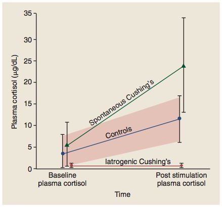
In normal dogs, administration of ACTH produces a rise in serum cortisol to values usually >10 μg/dl (>300 nmol/L). In contrast, dogs with Cushing’s syndrome (because of the increased thickness of the adrenal cortex) tend to have an exaggerated cortisol response to ACTH administration, with post-ACTH serum cortisol rising to concentrations >20 μg/dl (>600 nmol/L). Dogs with iatrogenic Cushing's have a blunted cortisol response to ACTH (see figure).

But what’s the percentage of dogs with Cushing’s disease that are actually diagnosed with this test? What’s the test sensitivity?

About half of dogs with cortisol-secreting adrenocortical tumors and about 80% of dogs with pituitary-dependent hyperadrenocorticism (PDH) show an exaggerated cortisol response to the ACTH stimulation test.

In clinical practice and reality, however, the sensitivity is actually worse than those statistics reveal. Although it is clear that over 20% of dogs with Cushing's syndrome have post-ACTH cortisol test results within the reference range (< 20 μg/dl for my laboratory), an additional 20-30% of dogs with Cushing's have test results described as "borderline" (serum cortisol concentrations >16 μg/dl but <24 μg/dl).

In effect, that means that only 50-60% of dogs with Cushing's disease will have serum cortisol responses that are clearly abnormal (> 25 μg/dl). In other word, this test is a poorly sensitive diagnostic test for Cushing’s syndrome. In most dogs, it would be more cost effective to turn to another test, such as the low-dose dexamethasone suppression test, as the first-line screening test.

Other disadvantages to using the ACTH stimulation test:

In addition to the ACTH stimulation test’s mediocre test sensitivity, there are 2 other reasons for choosing one of the other diagnostic tests over the ACTH stimulation test, including the following.
  1. First, of all, the cost of the preferred ACTH (cosyntropin; Cortrosyn) is high. I’ll discuss more about why you need to use cosyntropin in my next post (I’ll also offer my suggestions on how to extend the shelf-live of Cortrosyn to make it more cost effective).
  2. Secondly, this test cannot determine the underlying cause of Cushing's syndrome. In other words, a positive ACTH stimulation test doesn't tell you whether the dog has PDH or FAT, so additional testing will always be needed to determine that. It is a true screening test, and no features of the ACTH stimulation test result allow discrimination between PDH and FAT.
Are there any clinical situations where we should use the ACTH stimulation test?

Yes, there are definitely 3 situations in which ACTH stimulation testing would be indicated as a diagnostic or monitoring test for adrenal disease in dogs. The major indications for using the ACTH stimulation include the following:
  1. Best test to diagnose iatrogenic hyperadrenocorticism in dogs. If the dog has clinical and laboratory features consistent with Cushing’s syndrome (e.g., polyuria, polydipsia, polyphagia, pot-belly, truncal hair loss, high serum alkaline phosphatase) but has a recent history of glucocorticoid use, this is the test of choice. If the cortisol response to ACTH stimulation is low-normal or blunted in this dog, the diagnosis would be iatrogenic Cushing’s, rather than the naturally occurring disease. None of the other pituitary-adrenal function tests can make this differentiation.
  2. Best test to use for monitoring and adjusting mitotane or trilostane therapy. Again, this is the only pituitary-adrenal function test that can be used in this situation; none of the other pituitary-adrenal function are useful in monitoring drug treatment.
  3. “Gold Standard” test to diagnose spontaneous hypoadrenocorticism (Addison’s disease). This is true whether the dog has primary or secondary hypoadrenocorticism or typical or atypical disease; the ACTH stimulation test is the only test to use for this diagnosis. As you can see in the figure below (an Ad from the company that manufactures Cortrosyn), the main indication for ACTH as a diagnostic test is adrenocortical insufficiency, not Cushing's syndrome.
In general, there is a general rule for endocrine disease that is useful to remember when approaching endocrine hyperfunction vs. hypofunction:

In almost all cases, it is better to use stimulation tests to diagnose endocrine hypofunction, whereas suppression tests are best used for endocrine hyperfunction.

So with the ACTH stimulation, the 3 situations (listed above) where this test really has advantages over the other screening tests all include situations where the adrenal glands have been suppressed (iatrogenic steroid use, trilostane, mitotane) or destroyed (Addision’s disease)!

Monday, February 21, 2011

Q & A: Does this Dog have Cushing's Syndrome?

I recently ran a low-dose dexamethasone suppression test on a 11-year-old male neutered Maltese with clinical features of muscle loss, thinning coat, and a pot-bellied appearance.  His serum chemistry panel is completely normal except an alkaline phosphatase activity in the thousands.  Below are the results low-dose dexamethasone suppression test, but I am confused as to what to make of them.
  • Basal cortisol:  5.1 μg/dl (reference range = 1-4 μg/dl)
  • 5-hr post cortisol:  <0.7 μg/dl (reference range = <1.5 μg/dl)
  • 8-hr post cortisol:  1.9 μg/dl (reference range = <1.5 μg/dl)
Thanks in advance.


My Response:

The interpretation would be as follows: Slightly high basal cortisol, with complete suppression of cortisol at 5 hours, followed by escape from cortisol suppression at 8 hours.

The lack of suppression at the 8-hour testing period is diagnostic for hyperadrenocorticism. The fact that the dog suppressed so well at 4 hours tells us that this dog's Cushing's disease cannot be caused by an adrenal tumor (ie, these dogs would not suppress cortisol with either the low- or high-dose dexamethasone suppression test).

The pattern of cortisol suppression at 3-5 hours followed by escape from cortisol suppression at 8 hours, as shown here in this dog, is always diagnostic for pituitary-dependent hyperadrenocorticism.

Monday, January 24, 2011

How Do We Make Desmopressin Dose Adjustments in Dogs or Cats with Diabetes Insipidus?

Once a diagnosis of diabetes insipidus has been confirmed, the next step to start replacement treatment with desmopressin.

Initial treatment with desmopressin

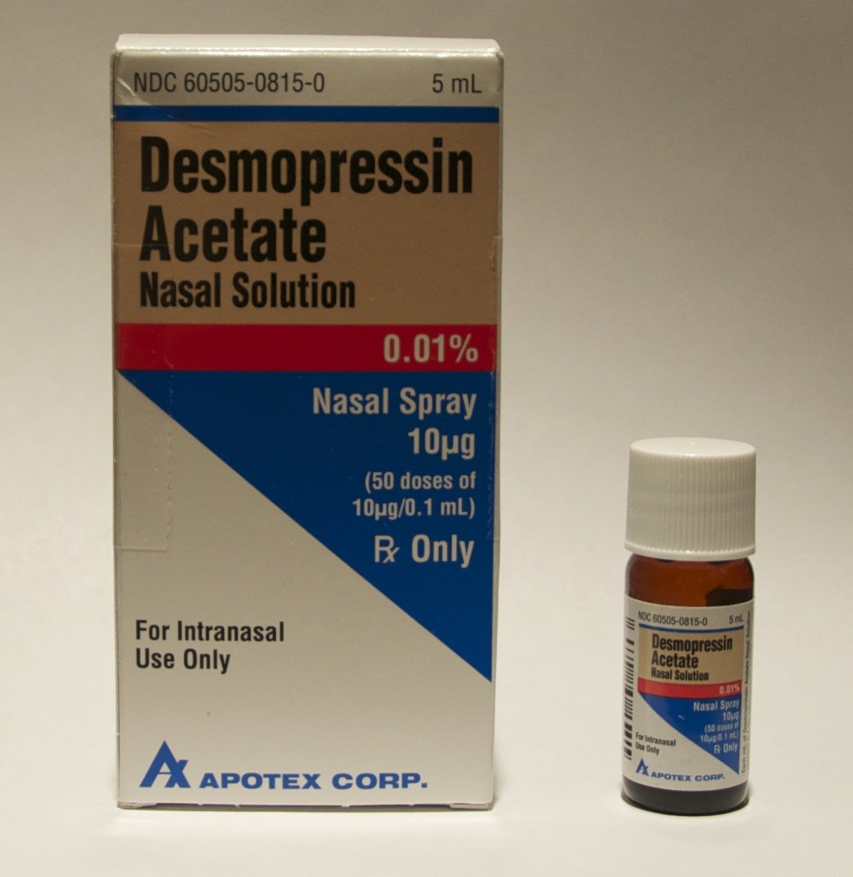
Recommended initial doses of desmopressin vary depending on the route it is being administered. In most cats and smaller dogs, 1 to 2 drops of the intranasal preparation administered once or twice daily are sufficient to control polyuria and polydipsia (see Table below).  Larger dogs may require up to 4 to 5 drops twice daily. Use of a tuberculin or insulin syringe allows for more accurate dosing. Application of desmopressin into the conjunctival sac may cause local irritation, as the solution is acidic. Some animals may object to the daily eye drops, making this route of administration ineffective.

With the subcutaneous route of administration, the initial recommended dose is 1.0 to 5.0 μg once or twice daily, depending on the size of the animals. If the nasal solution (100 µg /ml) were used for this purpose, one would inject only 0.01 to 0.05 ml (or 1 to 5 U with a U-100 insulin syringe). With the oral tablets, a starting dose of 0.05 mg to 0.2 mg (50 to 100 µg) once or twice daily is initiated.

Desmopressin dose adjustments

In dogs and cats with central diabetes insipidus, daily administration of desmopressin may completely eliminate polyuria and polydipsia. However, because of individual differences in absorption and metabolism, the dose required to achieve complete, around-the-clock control varies from patient to patient. The maximal effect of desmopressin occurs from 2 to 8 hours after administration, and the duration of action varies form 8 to 24 hours. Larger doses of the drug appear to both increase its antidiuretic effects and prolong its duration of action; however, expense can become a limiting factor for some owners.

No matter what route of administration is used, the daily dose should be gradually adjusted as needed to control signs of polydipsia and polyuria. The morning and evening doses can be adjusted separately if needed.

Adverse effects of desmopressin 

Desmopressin is relatively safe for use in animals with central diabetes insipidus. Adverse effects of desmopressin are uncommon, but overdosage can lead to fluid retention, hyponatremia, and decreased plasma osmolality. Although extremely rare, fluid intoxication associated with desmopressin overdosage can lead to CNS disturbances including depression, increased salivation, vomiting, ataxia, muscle tremors, coma and convulsions. In such instances, furosemide can be given to induce diuresis.

To avoid the potential problem of overdosage, it is recommended that animals not be allowed free access to water immediately after each dose of desmopressin, especially if severe polydipsia and polyuria have redeveloped. Without such short-term (1 to 2 hours) water restriction, the cat many consume excessive amounts of water that cannot be subsequently excreted, as the desmopressin is absorbed and has its peak antidiuretic effects on the renal tubules.

Cost of desmopressin

The principle drawback with the use of any of the desmopressin preparations in the treatment of central diabetes insipidus is the drug’s considerable expense.  The oral route of administration is the most expensive, while the subcutaneous route of administration (using the sterilized nasal solutions) is generally the most cost-effective.


Click on Table to enlarge.

Thursday, January 13, 2011

Diagnosis of Diabetes Insipidus: Is the Water Deprivation Test Necessary?

Several different diagnostic approaches can be used to confirm and distinguish central diabetes insipidus, nephrogenic diabetes insipidus and primary (psychogenic) polydipsia. The water deprivation test is generally considered by most authorities to be the best diagnostic test to differentiate between these disorders. However, the water deprivation test has many disadvantages, which include the following:
  • The test is very labor intensive
  • The test can be difficult to perform correctly
  • The test is quite unpleasant for the dog or cat
  • The test relies heavily on repeated emptying of the bladder
  • The test can lead to untoward complications
  • The test can lead to misdiagnosis in some animals

A simpler and more practical method of diagnosis that I recommended as an alternative to water deprivation testing is evaluation of the clinical response to a closely monitored therapeutic trial with the vasopressin analogue, desmopressin (DDAVP).

Use of the therapeutic trial with desmopressin as a diagnostic test 

This approach is less complicated and time consuming than the water deprivation test, and is certainly easier on the cat. The cost of the two approaches varies according to circumstances but is often comparable. Again, before a desmopressin trial is initiated, it is extremely important to rule out all other common causes of polyuria and polydipsia, limiting the differential diagnosis to central diabetes insipidus, primary nephrogenic diabetes insipidus, and primary (psychogenic) polydipsia. For further information, see my previous blog posts.

To perform the test, the owner should first measure the animal's 24-hour water intake for 2 to 3 days before desmopressin is initiated, allowing free-choice water intake. The dog or cat is then treated with therapeutic dosages of desmopressin (see our next post, What Drugs Do We Use to Treat Diabetes Insipidus).

For the purposes of this test, the desmopressin ideally is administered subcutaneously at the dosage of 1.0 to 4.0 μg twice daily for a period of 5 to 7 days. If subcutaneous injections cannot be given, administration of desmopressin by the conjunctival (1-5 drops twice daily) or oral routes (0.05-2.0 mg twice daily) can be used instead. During this treatment period, the owner should continue to measure the animal’s daily water intake and monitor the degree of urine output.

A dramatic reduction in water intake (>50% of pre-treatment measurements) and polyuria strongly suggests a diagnosis of central diabetes insipidus, whereas a lack of any reduction in polydipsia and polyuria is most consistent with primary nephrogenic diabetes insipidus. With more prolonged treatment, water consumption and urine output should completely normalize in dogs and cats with central diabetes insipidus.

In any older dog or cat that develops diabetes insipidus, one should consider pituitary imaging with computerized tomography or magnetic resonance imaging to exclude a pituitary mass. This is especially true if the affected animal has associated neurological signs.


Computerized tomography (CT) image of the brain of a cat with diabetes insipidus. Note the very large pituitary tumor invading the hypothalamus.