Saturday, January 28, 2012

Q & A: Blunted Cortisol Response to ACTH in a Dog with Suspected Addison's Disease


My patient is a 6.5-year old male neutered Belgian Malinois who has a history of weight loss and intermittent vomiting of 3-weeks duration.

A CBC and serum chemistry panel were both unremarkable, with normal serum sodium and potassium values. Abdominal ultrasonography was performed, which revealed a splenic mass. A splenectomy and liver biopsy were performed, which revealed a splenic hematoma and hepatopathy, thought to be secondary to Rimadyl (1). The drug has subsequently been discontinued with no improvement.

Today we did an ACTH stimulation test to rule out Addison's disease, using a dose of 2.2 U ACTH gel administered intramuscularly. The baseline cortisol value was 2.2 μg/dl (reference range, 1-4 μg/dl) and the 2-hour post-ACTH cortisol was 3.6 μg/dl (reference range, 6-20 μg/dl).

Is this subnormal cortisol response consistent with mild Addison's disease?

My Response:

This ACTH stimulation test completely rules out spontaneous hypoadrenocorticism (Addison's disease). Most dogs with primary, secondary, or atypical hypoadrenocorticism will have a low basal cortisol concentration (< 1 μg/dl) and all will have a baseline cortisol value that is less than 2 μg/dl (2-4).

After ACTH stimulation, dogs with hypoadrenocorticism tend not to have any cortisol response to ACTH stimulation, and post-ACTH cortisol values should never be greater than 2.5-3.0 μg/dl (see Figure below)

In many dogs, the blunted cortisol response, as seen in this dog, is either due to prior administration of glucocorticoid sometime in the last few weeks. However, in this dog, the likely cause for the subnormal cortisol response is the compounded ACTH preparation that you used.

In a study performed a few years ago (5), normal dogs were tested with cosyntropin (Cortrosyn, 5 μg/kg, IV) and 4 different compounded forms of ACTH (all manufacturers recommend a dose of 2.2 U/kg, IM).  For all 5 of the ACTH stimulation tests, samples for cortisol analysis were collected before and 1 and 2 hours after ACTH injection.

After administration of the 4 forms of compounded ACTH, the time to reach peak cortisol concentrations varied, with some dogs showing the maximal cortisol value at 1 hour and others having peak cortisol concentrations at 2 hours.With 2 of the 4 products, serum cortisol concentration returned to baseline values by 2 hours in many of the dogs.

Thus, due to variability in duration of cortisol response after injection of the compounded ACTH products, the investigators recommend that we need to collect several post-ACTH samples for cortisol analysis after administration of a compounded ACTH preparation to ensure that we detect the peak cortisol response. At the minimum, we recommend collecting samples at 1 and 2 hours following injection.

Because of this variability in cortisol stimulation, I'm not a fan of using compounded ACTH preparations. I would rather use low-dose Cortrosyn (6) and collect only one post-ACTH blood sample at 1-hour and be assured that will produce the maximal cortisol response at that time.

References:
  1. MacPhail CM, Lappin MR, Meyer DJ, et al. Hepatocellular toxicosis associated with administration of carprofen in 21 dogs. Journal of the American Veterinary Medical Association 1998; 212: 1895-1901.
  2. Lifton SJ, King LG, Zerbe CA. Glucocorticoid deficient hypoadrenocorticism in dogs: 18 cases (1986-1995). Journal of the American Veterinary Medical Association 1996;15;209:2076-2081. 
  3. Cook AK, Bond KG. Evaluation of the use of baseline cortisol concentration as a monitoring tool for dogs receiving trilostane as a treatment for hyperadrenocorticism. Journal of the American Veterinary Medical Association 2010;237:801-805.
  4. Klein SC, Peterson ME. Canine hypoadrenocorticism: part II. Canadian Veterinary Journal 2010;51:179-184.
  5. Kemppainen RJ, Behrend EN, Busch KA. Use of compounded adrenocorticotropic hormone (ACTH) for adrenal function testing in dogs. Journal of the American Animal Hospital Association 2005;41:368-372. 
  6. Martin LG, Behrend EN, Mealey KL, et al.  Effect of low doses of cosyntropin on serum cortisol concentrations in clinically normal dogs. Journal of the American Veterinary Medical Association 2007;230:555-560. 

Thursday, January 26, 2012

Q & A: Diabetes Insipidus in a Hypothyroid Golden Retriever

I have an interesting situation with a dog with polyuria and polydipsia (PU/PD) that I have not seen before, and wonder if you can offer any insight.

My patient is a 7-year old, male neutered Golden Retriever-mix weighing 69 pounds who was diagnosed as being hypothyroid and has been on L-thyroxine replacement (Soloxine) for at least 5 years. He was recently diagnosed with diabetes insipidus (DI) because of signs of severe PU/PD and low urine specific gravity. The dog was examined by a neurologist, who found no neurological deficits. On MRI examination, the dog had no abnormal findings to explain the onset of DI (i.e, no hypothalamic or pituitary lesions or masses). 

He is now on Soloxine at a dosage of 0.7 mg twice daily (there has been no change in L-T4 dose for last year). For the DI, we have him on 0.01% desmopressin, with drops administered twice daily into the conjunctival sac (1 drop in morning and 2 drops in evening).

His owner reports he has been doing great in the last 3 to 4 months since he added the desmopressin. Recently, however, we rechecked a 6-hour post-pill serum T4 concentration and found that the value was high at 5.5 μg/dl (reference range, 1-4 μg/dl).

My questions are:
  1. Can this be due to an interaction of desmopressin and L-thyroxine?
  2. Is this a result of correcting/properly treating his diabetes insipidus and is this common?
  3. Do we need to take any special factors into consideration with dosing either drug, or just lower his Soloxine as we would in other hypothyroid dogs getting routine T4 rechecks for proper dosing?
  4. Is this just a coincidence and no known correlation?
My Response:

The main sign of hyperthyroidism in dogs is PU/PD so I'm be a bit worried that the thyroid hormone supplementation is causing the signs (1-4). You could drop the dose down a bit to see if that helps the PU/PD. After a week or so on the reduced dose of L-T4, you could try stopping the desmopressin for a couple of days and see if the PU/PD returns.

I don't know of any problems with concurrent use of desmopressin and thyroid hormone. But it is somewhat unlikely that a 7-year old dog would develop DI without the presence of any obvious pituitary or CNS pathology (1,2,4).

Many dogs with undefined PU/PD will respond to desmopressin but most of those dogs do not have DI. I'm not saying that we shouldn't use the desmopressin to control the signs, but DI is very unlikely.

Follow-up Question:

Thank you for your ideas, Dr. Peterson.

Do you mean that perhaps he was being overtreated for his hypothyroidism thus making him hyperthyroid leading to the PU/PD that initially caused us to diagnose the diabetes insipidus? I'm pretty sure that his T4 was in the normal range at that time. And now when we just tested him and discovered the T4 high, he is no longer PU/PD (since being on the desmopressin).

That is interesting that even dogs who do not truly have DI will respond to desmopressin. Will desmopressin eventually stop helping though? Or would you just keep him on it as long as it controls the severe PU/PD?

My Response:

Remember that when we are doing a post-pill T4, we are only doing a single spot check. That doesn't really indicate that is happening throughout the day.

In addition, the finding of a serum T4 value within the lab's reference range does not indicate what that particular dog's individual T4 normal range actually is — in other words, some dogs may be perfectly euthyroid having a serum T4 value of 4.5 μg/dl, whereas others may be clinically hyperthyroid with a serum T4 of 3.5 μg/dl.

And yes, most dogs that respond to desmopressin do not really have DI. As long as you and the client realize that, this is a pretty safe drug and it can be used to control PU/PD for long periods (and maybe forever).

Again, I'd still try cutting back on the L-T4 dose first -- why give desmopressin if we can figure out the real cause of the PU/PD?

References:
  1. Peterson ME, Nichols R: Investigation of polyuria and polydipsia, In: Mooney C.T., Peterson M.E. (eds), Manual of Canine and Feline Endocrinology (Third Ed), Quedgeley, Gloucester, British Small Animal Veterinary Association, pp 16-25, 2004.
  2. Randolph JF, Nichols R, Peterson ME: Diseases of the hypothalamus and pituitary. In: Birchard SJ, Sherding RG (eds): Manual of Small Animal Practice (Third Edition), Philadelphia, Saunders Elsevier, pp 398-408, 2006.
  3. Peterson ME: Hyperthyroidism and thyroid tumor in dogs. In: Melian C, Perez Alenza MD, Peterson ME, Diaz M, Kooistra H (eds): Manual de Endocrinología en Pequeños Animales (Manual of Small Animal Endocrinology). Multimedica, Barcelona, Spain, 2008, pp 113-125.
  4. Nichols R, Peterson ME: Investigation of polyuria and polydipsia, In: Mooney C.T., Peterson M.E. (eds), Manual of Canine and Feline Endocrinology (Fourth Ed), Quedgeley, Gloucester, British Small Animal Veterinary Association, 2012; 215-220.

Sunday, January 22, 2012

Q & A: Diagnosing Cushing's Disease in Dogs with Diabetes Mellitus

Rex is an 8-year-old male Schnoodle who presented to us a month ago with owner complaints of polyuria and polydipsia (PU/PD) and polyphagia. On physical examination, his abdomen was a bit pendulous, with a body condition score of 7/9 (body weight, 8.5 kg). Marked hepatomegaly was found, which was verified by abdominal radiography. The physical examination was otherwise unremarkable, with no other signs of Cushing's syndrome (e.g., no hair loss, muscle atrophy, comedomes, calcinosis cutis).

We submitted a complete blood count, serum chemistry panel, complete urinalysis, and urine culture to the lab that day, which showed the following abnormalities:

Serum Chemistry Panel:
  • Alkaline phosphatase: 1061 IU/L (reference range, 5-131)
  • GGT: 18 IU/L (reference range, 1-12)
  • Glucose: 649 mg/dl (reference range, 70-138)
  • Triglyceride 414 mg/dL (reference range, 29-291)
Urinalysis and Culture
  • Specific gravity: 1.029
  • Glucosuria: 3+
  • Ketones: Negative  
  • Urine culture: negative
Based on the hyperglycemia and glycosuria we diagnosed diabetes mellitus and started Rex on NPH insulin (Humulin-N) at the dosage of 4 U BID (0.5 U/kg/injection). He responded to insulin with a moderate decrease in PU/PD, but the thirst remained excessive.

We did an serial blood glucose cure 2 weeks later. Results showed marked and persistant hyperglycemia (> 400 mg/dl) throughout the day. Based upon the glucose curve, we diagnosed insulin resistance and did a low-dose dexamethasone response test. Results of that test were positive, and indicate pituitary-dependent Cushing's disease (see below):
  • Basal cortisol: 5.8 μg/dl (reference range, 1.0-5.0 μg/dl)
  • 4-hr post-cortisol: 0.9 μg/dl (reference range, <1.4 μg/dl)
  • 8-hr post-cortisol:  5.7 μg/dl (reference range, <1.4 μg/dl)
I'd like to start Rex on trilostane (Vetoryl, Dechra Animal Health) for the Cushing's disease and insulin resistance. Am I on the right course with this dog?

My Response:

The problem with diabetic dogs is that it's very difficult to make a diagnosis of Cushing's with certainty unless we see cutaneous changes. PU/PD, polyphagia, and high liver values all could be secondary to the diabetes. False-positive test results on a LDDST are very common in dogs with nonadrenal illness, such as diabetes (1-3).

Does he have any cutaneous signs (hair loss, etc)? His insulin dose isn't high enough to say that he has insulin resistance. I'd recommend that you continue to monitor Rex closely an slowly raise his NPH dose to see if that helps control his signs. Because it can be very difficult to be sure in these diabetic dogs, sometimes observation and monitoring is the best course.

Follow-up:

Rex's coat looks pretty normal right now. We'll try raising his insulin dose before assuming that Cushing's disease is his underlying problem. Two more questions:
  1. At what insulin dose do we get concerned about resistance?
  2. Could diabetes explain the pot belly and marked hepatomegaly?
My Response:

We define insulin resistance as doses greater than 2.2 U/kg/injection to control hyperglycemia (4), so we aren't even close to the doses required to diagnose resistance.

And yes, diabetic dogs can get marked hepatomegaly secondary to fat accumulation in the liver. That can lead to a mild-moderate "pot bellied" appearance.

Because it can be very difficult to make a diagnosis of Cushing's syndrome in dogs with diabetes, observation and monitoring is the best course in many of these patients. If Cushing's disease is present, it will be progressive and other signs will develop to make the diagnosis easier to confirm.

References:
  1. Kaplan AJ, Peterson ME, Kemppainen RJ. Effects of disease on the results of diagnostic tests for use in detecting hyperadrenocorticism in dogs. Journal of the American Veterinary Medical Association 1995;207:445-451.
  2. Kaplan A, Peterson ME. Effects of nonadrenal disease on adrenal function tests In: Bonagura JD (ed): Current Veterinary Therapy XIII. Philadelphia, WB Saunders Co., 2000; pp 362-363.
  3. Melián C, M. Pérez-Alenza, D, Peterson ME. Hyperadrenocorticism in dogs, In: Ettinger SJ (ed): Textbook of Veterinary Internal Medicine: Diseases of the Dog and Cat (Seventh Edition). Philadelphia, Saunders Elsevier, 2010; pp. 1816-1840.
  4. Peterson ME. The difficult diabetic: Acromegaly, Cushing’s, and other causes of insulin resistance. North American Veterinary Conference (NAVC) Conference 2012: Small Animal & Exotics Proceedings. pp. 873-879.

Thursday, January 19, 2012

Believe The Hype? Hill's Latest Marketing Slogan for the y/d Diet

I attended the North American Veterinary Conference (NAVC) in Orland Florida this week, where I presented a number of lectures on a variety of endocrine topics (complete list of topics on my website). 

One lecture that I had agreed to present 9 months ago was part of a Hill's seminar on diagnosis and treatment of feline hyperthyroidism. The topic that I had agreed to present (which I did present last Monday morning on January 16th), was entitled "Feline Hyperthyroidism: Risk Factors and Diagnosis." This was arranged before Hill's iodine deficient y/d diet had been released, so the intense marketing campaign that we have seen over the last 3 months had not yet begun.


The day before I traveled to the NAVC conference, I received an email from Hill's with the above ad, promoting the hyperthyroid seminar that I would soon be a part of — to be honest, I was shocked and rather embarrassed that I was to be a part of this y/d promotion.

Once I arrived in Orlando, the slogan, Believe The Hype was posted as a prominent banner almost everywhere that one looked. Again, I was feeling more and more uncomfortable that I was to be included as part of this "hype,"especially since I'm not convinced that this diet is even safe to use, based on the evidence.  (To see my past posts which discuss the y/d diet, click here.)

Remember that we have yet to see any referred scientific publications with the complete data that answer all of our questions. The Hill's research team certainly hasn't responded to most of my questions that I've brought up about y/d in my blogs.

Defining the Term, Hype

After thinking about "Believe The Hype" banner and discussing the matter over dinner with friends, someone asked the obvious question —What does the word hype really mean, anyway?

Well, according to the Merriam-Webster Dictionary, the word, when used as a noun as it is in the slogan, has two possible meanings:
  1. deception, put-on
  2. publicity; especially: promotional publicity of an extravagant or contrived kind 
And according to an online dictionary (dictionary.com), the word hype has three meanings, all similar to those found in the Merriam-Webster  source:
  1. exaggerated publicity; hoopla
  2. an ingenious or questionable claim, method, etc., used in advertising, promotion, or publicity to intensify the effect
  3. a swindle, deception, or trick
So What Is Hill's Implying with the Believe The Hype Slogan?

At face value, is the marketing division at Hill's actually asking us to believe that the information they have provided about the y/d diet is all hype? Remember that the word is defined as exaggerated, ingenious or questionable claims, put-ons, or tricks!

Interesting thought, isn't it.

Personally, I find the slogan to be "over-hyped" — unnecessarily exaggerated to the point of overkill. Why don't they present the hard evidence, answer the real questions that we all have, and forget this foolishness?

My Bottom Line

We all need to remember that this marketing campaign for the Hill's y/d diet, as well as the marketing ads for the multitudes of products that we get bombarded with every day, is all about hype.

As veterinarians and advocates for our feline patients, our job is to dig below this exaggerated hype and find the real science (if any) behind this sensational publicity. In the case of y/d, Hill's has created much more hype than usual or really necessary (at least in my opinion).  If half of this money spent on this marketing campaign would be directed toward research on feline hyperthyroidism, we may be able to find the cure for this disease!

I know that it is tempting to get taken in with all marketing of new products, especially y/d. My goal in writing these blog posts, the way I see it, is to try to bring everyone back to earth and make the practicing veterinarian think about the possible long-term implications of feeding y/d to cats. That's what I have tried to do over the past few weeks and will continue to do as needed in the future.

Hill's y/d diet is different than all of the other commercial pet foods on the market because they are promoting y/d as a treatment for a disease — in other words, Hill's is marketing y/d more like a drug than a diet per se.  In that regard, I believe that they need to do the studies to ensure that this diet is safe, as well as effective, just as any pharmaceutical company would have to do for FDA approval.  From what I've heard, however, no further safety studies are planned simply because they are not required to do them.

But is that the "right" thing to do, just because the diet isn't regulated as a drug by the FDA? Or is it all about selling as much cat food as possible to keep the shareholders happy?

Tuesday, January 10, 2012

Q & A: If Nuclear Accidents Can Lead to Thyroid Cancer, Why Doesn't the I-131 Treatment?

Thanks for your great post on radioprotective therapies (see Radiation Protection after a Nuclear Accident).

But I'm confused. If radioidine-131 released into the atmosphere during an accident can result in a dramatic increase of thyroid cancer in children, why don't people or cats treated with I-131 for hyperthyroidism develop thyroid cancers?

My Response:

Good question. After a nuclear accident, people and animals will get exposed to varying amounts of radioiodine as I-131, as well as many other radioisotopes including cesium-137, strontium-90, and plutonium-241 (1). Some will receive a small dose to their thyroid, whereas others will get more, but it's generally not enough to destroy the thyroid — it "tickles" the thyroid and causes DNA damage, which can lead to thyroid tumors (2).  The exposure also tends to happen over a period of a few days to weeks, not all at once.

When we treat a patient (human, cat, dog) with radioiodine, we are delivering a huge, single dose of radiodine to the thyroid. This destroys the hyperfunctional adenoma/carcinoma. It tends not to be taken up by the normal, suppressed tissue. It is unclear why the remaining normal thyroid tissue doesn't develop cancer, but it appears related to the large, single dose of radioiodine administered, as opposed to chronic exposure of lower levels of radiation. It has been reported that spreading the total I-131 dose over time (from a few days to a few weeks or longer) may lower risk of thyroid cancer, probably due to the opportunity for cellular repair mechanisms to operate (3).

Infants and children have the peak risk as the result of the increased radiation sensitivity of their thyroid glands. As a result, most human patients that develop thyroid cancer after exposure to radioiodine are babies and young children (4-6), whereas adult individuals are at a lesser risk. This fact probably also contributes to why we don't see thyroid cancer developing in adult human or feline patients treated with radioiodine.

The latest study in Hiroshima and Nagasaki Atomic Bomb survivors in Japan has indicated that a biological effect from a single brief external exposure to ionizing radiation nearly 60 years in the past still occurs and can be detected (7). In childhood, once exposed even to low doses of ionizing radiation, either externally or internally, the cancer-prone cell damage within the thyroid gland can be preserved for a long time.

What Can We Do in an Nuclear Emergency?

Since children are at the highest risk to exposure to radioactive iodine, potassium iodide should be available to all children (8). Also, because of the risk to the developing fetus, pregnant women should also take potassium iodide in the event of a nuclear accident. Compared to children and the fetus, adults are at a lower risk but still would benefit from thyroid blockage with potassium iodide.

Iosat Tablets, one FDA approved KI preparation
Potassium iodide pills (sometimes abbreviated as KI: the K stands for potassium, the I for iodine) don’t prevent radioactive iodine from entering the body. But these pills do keep the radioiodine from accumulating in the thyroid gland. By flooding the body with non-radioactive iodine, the pills keep the gland from absorbing the radioactive iodine. Here is how the CDC fact sheet summed it up (9):

Because KI contains so much stable iodine, the thyroid gland becomes “full” and cannot absorb any more iodine—either stable or radioactive—for the next 24 hours.
ThyroShield and ThyroSafe, the 2 other FDA approved KI preparations

What Potassium Iodide Preparations Are Available?

The potassium iodide (KI) products approved by the Food and Drug Administration (FDA) include the following (10):
Properly packaged, potassium iodide’s shelf life is at least 5 years and possibly as long as 11 years. If you take a very old pill, it may not work but won't hurt you or do any harm.

What are the Recommended Potassium Iodide Doses?

For humans, the FDA recommends the following doses based on the patient's age:
  • 0—1 months ........................................ 15 mg
  • 1 months—3 years ............................30-35 mg
  • 3—12 years ...........................................65 mg
  • >12 years ............................................130 mg
As I discussed in my last post, the recommended dose of potassium iodide for a dog 1.4 mg/kg (11). In other words, a 20-kg dog would receive 25-30 mg, whereas a small dog or cat would receive about 5-10 mg.  The easiest way to prepare a 15-mg dose for a newborn is to dissolve a 130-mg pill in 8 oz of a clear liquid and feed the newborn 1 oz of the liquid. Similar measures could easily be used in dogs and cats.

A single dose of KI protects the thyroid gland for up to 24 hours. A one-time dose at the levels recommended by the FDA is usually all that is needed to protect the thyroid gland. In the rare instance that radioiodine is present in the environment for longer than 24 hours, continuing the dose of KI every 24 hours for a few days would be needed.

References:
  1. Report of the Chernobyl Forum Expert Group. Environmental Consequences of the Chernobyl Accident and their Remediation: Twenty Years of Experience. International Atomic Energy Agency, Vienna, 2006. 
  2. Awa A. Analysis of chromosome aberrations in atomic bomb survivors for dose assessment: studies at the Radiation Effects Research Foundation from 1968 to 1993. Stem Cells. 1997;15 Suppl 2:163-73.
  3. Ron E, Lubin JH, Shore RE et al. Thyroid cancer after exposure to external radiation; a pooled analysis of seven studies. Radiation Research 1995;141:256-277. 
  4. Jacob P, Kenigsberg Y, Zvonova I, et al. Childhood exposure due to the Chernobyl accident and thyroid cancer risk in contaminated areas of Belarus and Russia. British Journal of Cancer 1999;80:1461-1469. 
  5. Ivanov VK, Gorski AI, Maksiutov MA, et al. Thyroid cancer incidence among adolescents and adults in Bryansk region of Russia following the Chernobyl accident. Health Physics 2003;84: 46-60. 
  6. Cardis E, Kesminiene A, Ivanov V, et al. Risk of thyroid cancer after exposure to I-131 in childhood. Journal of the National Cancer Institute 2005;97:724-732. 
  7. Imaizumi M, Usa T, Tominaga T, et al. Radiation dose-response for thyroid nodules and autoimmune thyroid diseases in Hiroshima and Nagasaki Atomic Bomb Survivors 55-58 years after radiation exposure. Journal of the American Medical Association 2006;295:1011-1022. 
  8. National Research Council. Distribution and  administration of potassium iodide in the event of a nuclear incident, Washington, DC: National Academies Press, 2004.
  9. Center for Disease Control and Prevention website. Potassium iodide factsheet
  10. FDA website. Frequently asked questions on potassium iodide (KI). www.fda.gov
  11. Ribela MT, Marone MM, Bartolini P. Use of radioiodine urinalysis for effective thyroid blocking in the first few hours post exposure. Health Physics 1999;76:11-16.

Thursday, January 5, 2012

Paper Review: Radiation Protection after a Nuclear Accident

Scientific Paper Review— Facing the Nuclear Threat: Thyroid Blocking Revisited

Hänscheid H, Reiners C, Goulko G, Luster M, Schneider-Ludorff M, Buck AK, and Lassmann M. Facing the nuclear threat: thyroid blocking revisited. Journal of Clinical Endocrinology and Metabolism 2011;96:3511-3516.

Two common preparations of Potassium Iodide available in the US

Summary of Paper

Background:
Radioiodine-131 is a major fission product released into the atmosphere after a nuclear accident and results in contamination of water and soil. It may be ingested through food and water that is contaminated with 131-iodide. The most recent accident, of course, was the nuclear reactor damage from the Tsunami in Japan.

Radioidine-131 from the Chernobyl accident resulted in a dramatic increase of  thyroid cancer in children who were exposed to the radiation. To prevent thyroid  cancer from thyroid uptake of 131-iodide, the World Health Organization and the U.S. National Research Council have recommended that the potentially exposed population be given tablets containing 100 mg of iodide as potassium iodide to block the uptake of the radioiodide (1).

Potassium iodide is the same form of iodine used to iodize table salt. Potassium iodide floods the thyroid with iodine, thus preventing radioactive iodine from being absorbed (2). Taking potassium iodide immediately after a nuclear accident appears to lessen the risk of developing thyroid cancer.

Sodium perchlorate is a chemical that causes the thyroid to release any iodine that is stored in the thyroid cells (3). Thus, taking sodium perchlorate after a nuclear accident may cause the thyroid to release any radioactive iodine that the thyroid cells have already taken up.

This study was done to see if taking sodium perchlorate might also lower the chance of thyroid cancer after a nuclear accident. To that end, the effectiveness of sodium perchlorate as a blocking agent was compared with the use of potassium iodide in patients exposed to radioactive iodine.

Methods:
Twenty-seven healthy euthyroid subjects with a mean age of 25 years participated in 48 studies of 123-I kinetics in the thyroid. None of these patients were known to have any thyroid disease.

Each volunteer received a small tracer dose of radioactive iodine (I-123), then different amounts of either potassium iodide or sodium perchlorate. The volunteers then had a radioactive iodine uptake (RAIU) test to see how long the radiation stayed in the thyroid.

Results:
The authors found that 100 mg of either potassium iodide or sodium perchlorate was able to reduce the thyroid absorbed dose of radioactive iodine by almost 90%.

Iodine kinetics were significantly faster in subjects younger than 25 years of age, as compared with those older than 25 years. The time of intervention to achieve a 50% dose reduction was 2.4 hours fort he person with the fastest kinetics and 9.2 hours for the subject with the slowest iodine kinetics.

Conclusions: 
At a time of a nuclear disaster, shielding from radiation by distance, staying indoors, and taking all other measures to avoid exposure to radiation are important. Public authorities must act quickly to distribute iodide tablets to the population who may be exposed in order to prevent thyroid uptake of 131-I that has strong beta radiation, which can induce DNA damage and result in thyroid cancer (1). The data of this paper show that the best blockade occurs when the blocking agent is given before the patient is exposed to radioiodine.

Currently, potassium iodide is the main compound to take after a nuclear accident and is stockpiled in areas that have nuclear power plants in the United States. The results suggest that both potassium iodide and sodium perchlorate lower radioactive iodine levels to the thyroid after a nuclear accident. Both agents are relatively safe to take as a single dose following a nuclear accident.

Since thyroid effects of low levels of perchlorate in the U.S. environment are controversial, however, it is unlikely that it will be made available to the general U.S. public following a nuclear accident. However, it is another compound that can be used in this situation.

Current guidelines for blocking 131- I uptake are adequate for older individuals but probably overestimate the efficacy of blocking in young people, who have faster kinetics. Perchlorate may be used for thyroid blocking as an alternative for individuals with iodine hypersensitivity or those at risk for iodine-induced hyperthyroidism. The conclusion that thyroid iodine kinetics vary among individuals is not novel, but the point that younger people have faster turnover and may need a larger blocking dose given at an earlier time is important.

Implications of the Study (including Protecting our Dogs and Cats)

What about our companion animals? The iodine kinetics for both dogs and cats tends to be more rapid than in human patients, which could suggest that larger doses of potassium iodide or perchlorate might be needed in case of an emergency.

In one study of normal dogs, the blocking action of both potassium iodide and potassium perchlorate was about 90%, similar to the findings of this present study (5). Potassium iodide was chosen for its limited side effects and more universal utilization. In that canine study, results indicated that 25 mg of potassium iodide is the ideal amount to be administered to the dog. This corresponds to approximately 100 mg for a 70 kg human being (i.e., a dose of 1.4 mg/kg).

For more information about use of potassium iodide in dogs and cats, see my related post on "Radiation Toxicity, Potassium Iodide, and Our Pets."

References:
  1. National Research Council. Distribution and  administration of potassium iodide in the event of a nuclear incident, Washington, DC: National Academies Press, 2004.
  2. Sternthal E, Lipworth L, Stanley B, Abreau C, Fang SL, Braverman LE. Suppression of thyroid radioiodine uptake by various doses of stable iodide. New England Journal of Medicine 1980;303:1083-1088.
  3. Greer MA, Goodman G, Pleus RC, Greer SE. Health effects assessment for environmental perchlorate contamination: the dose response for inhibition of thyroidal radioiodine uptake in humans. Environmental Health Perspectives 2002;110:927-937.
  4. Ribela MT, Marone MM, Bartolini P. Use of radioiodine urinalysis for effective thyroid blocking in the first few hours post exposure. Health Physics 1999;76:11-16.

Sunday, January 1, 2012

Q & A: Triiodothyronine (T3) Suppression Test Protocol For Cats

What form would Liothyronine sodium be given for a T3 suppression test?

What testing protocol do you use? Can you explain how you interprete the test results?

My Response:

Liothyronine sodium is the L-isomer of triiodothyronine (T3). I have always used tablets that contain 25 μg. The common trade name for L-T3 is Cytomel (Figure 1).

Protocol for T3 suppression test in cats

The test protocol that I developed over 20 years ago and still use is as follows (1):
Figure 1: Cytomel
  1. One day 1, draw a blood sample is drawn for determination of baseline serum concentrations of total T4 and T3. This serum sample is not yet submitted to the laboratory but kept refrigerated (or frozen) until day 4.
  2. Owners are instructed to give 7 doses of a T3 pill (liothyronine sodium; Cytomel) to their cat, beginning on the following morning.
  3. On day 2 and 3, the owners administer the liothyronine at a dosage of 25 µg every 8 hours for 2 days (6 doses).
  4. On the morning of day 4, a seventh 25-µg dose of liothyronine is given and the cat returned to the veterinary clinic within 2 to 4 hours.
  5. The veterinarian again draws a blood sample for serum T4 and T3 determinations.
  6. Both the basal (day 1) and post-liothyronine (day 4) serum samples are submitted to the laboratory together to eliminate the effect of between assay variation in hormone concentrations.
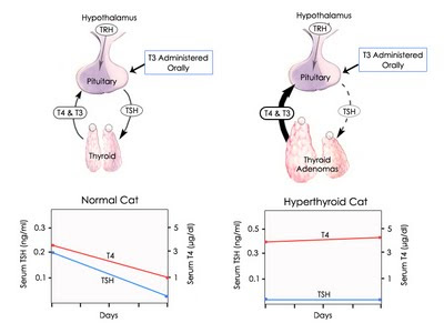
Figure 2: T3 suppression tests in normal cats (left) & cats with hyperthyroidism (right)
Interpretation of results of T3 suppression test

Regarding interpretation of T3 suppression test results, I find that the absolute serum T4 concentration after liothyronine administration is the best means of distinguishing hyperthyroid cats from normal cats or cats with nonthyroidal disease (1-5).

Cats with hyperthyroidism have post-liothyronine serum T4 values greater than 20 nmol/L (greater than 1.5 μg/dl), whereas normal cats and cats with nonthyroidal disease have T4 values less than 20 nmol/L (Figure 2). There may be a great deal of overlap of the per cent decrease in serum T4 concentrations after liothyronine administration between the three groups of cats, but suppression of 50 per cent or more only occurs in cats without hyperthyroidism.

Serum T3 concentrations, as part of the T3 suppression test, are not useful in the diagnosis of hyperthyroidism per se. However, these basal and post-liothyronine serum T3 determinations can be used to monitor owner compliance with giving the drug. If inadequate T4 suppression is found, but serum T3 values do not increase after treatment with liothyronine, problems with owner compliance should be suspected and the test result considered questionable.

Disadvantages of the T3 suppression test

Overall, the T3 suppression test is very useful for diagnosis of mild hyperthyroidism in cats, but the test does come with disadvantages:
  • T3 suppression testing is a relatively long test (3 days)
  • Owners are required to give multiple doses of liothyronine
  • Cats must swallow the tablets if the test is going to be meaningful.
  • If the liothyronine is not administered properly or the cat does not swallow the liothyronine tablet, circulating T3 concentrations will not rise to decrease pituitary TSH secretion (Figure 2), and the serum T4 value will not be suppressed, even if the pituitary-thyroid axis is normal. Failure of a cat to ingest the liothyronine could result in a false-positive diagnosis of hyperthyroidism in a normal cat or cat with nonthyroidal disease.

References:
  1. Peterson ME, Graves TK, Gamble DA: Triiodothyronine (T3) suppression test. An aid in the diagnosis of mild hyperthyroidism in cats. Journal of Veterinary Internal Medicine 1990;4:233-238.
  2. Peterson ME. Diagnostic tests for hyperthyroidism in cats. Clinical Techniques in Small Animal Practice 2006;21:2-9.
  3. Peterson ME: Diagnostic testing for feline hyper- and hypothyroidism. Proceedings of the 2011 American College of Veterinary Internal Medicine (ACVIM) Forum. pp. 95-97, 2011
  4. Peterson ME: Hyperthyroidism in cats, In: Rand, J (ed), Clinical Endocrinology of Companion Animals. New York, Wiley-Blackwell, in press.
  5. Mooney CT, Peterson ME: Feline hyperthyroidism, In: Mooney CT, Peterson ME (eds), Manual of Canine and Feline Endocrinology (Fourth Ed), Quedgeley, Gloucester, British Small Animal Veterinary Association, 2012; in press.